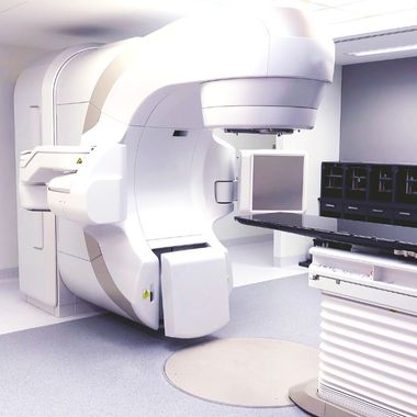

What is Radiology?
Radiology is a branch of medicine that uses imaging of the body's internal structures to diagnose and treat diseases. Radiology uses a variety of imaging techniques to diagnose, monitor, and plan treatment for diseases. These techniques include X-rays, computed tomography (CT), and ultrasonography (US).
X-rays (Roentgen): Used to evaluate bone fractures, joint problems, and some internal organ diseases.
Computed Tomography (CT): Provides detailed cross-sectional images and is used for detailed examination of internal organs, bones, and soft tissues.
Ultrasound (US): A radiation-free method used for examination of soft tissues, organs, and fetuses.
Bones and Joints: Fractures, dislocations, joint diseases, arthritis, osteoporosis.
Respiratory System: Lung diseases, pneumonia, tumors, pulmonary embolism.
Digestive System: Stomach and intestinal diseases, liver diseases, gallbladder stones.
Urinary System: Kidney stones, urinary tract infections, prostate problems.
Cardiovascular System: Heart diseases, vascular occlusions, aneurysms.
Nervous System: Brain and spinal cord diseases, stroke, tumors.
Women's Health: Breast tumors, ovarian cysts, pregnancy and birth processes.
Child Health: Congenital abnormalities, growth problems.
Breast Cancer: Screening and diagnosis with mammography and ultrasound.
Lung Cancer: Diagnosis and staging with CT and PET-CT.
Colorectal Cancer: Screening and evaluation with colonoscopy and CT.
Prostate Cancer: Evaluation and biopsy guidance with MRI and ultrasound.
Coronary Artery Disease: Evaluation of vascular structures with CT angiography.
Heart Failure: Evaluation of heart functions with ECHO (Echocardiography).
Aortic Aneurysm: Diagnosis and monitoring with CT and MRI.
Fractures and Dislocations: Diagnosis and treatment planning with X-ray.
Arthritis: Evaluation of joint changes with X-ray and MRI.
Low Back and Neck Pain: Diagnosis of spine and disc problems with MRI.
Stroke: Evaluation of brain tissue with CT and MRI.
Multiple Sclerosis (MS): Monitoring brain and spinal cord lesions with MRI.
Headaches: Investigating the causes of headaches with MRI and CT.
COPD (Chronic Obstructive Pulmonary Disease): Evaluation of lung changes with CT. Pneumonia: Diagnosis of lung infections with X-ray and CT.
Liver Diseases: Evaluation of the liver with CT, MRI and ultrasound.
Gallbladder Stones: Diagnosis with ultrasound.
Pancreatic Diseases: Evaluation of the pancreas with CT and MRI.
Kidney Stones: Diagnosis with ultrasound and CT. Urinary Tract Infections: Evaluation with ultrasound and radiological examinations.
Thyroid Diseases: Thyroid evaluation with ultrasound and scintigraphy (nuclear medicine).
Trauma: Evaluation of injuries resulting from trauma with X-ray and CT. Emergencies: Use of various imaging modalities for rapid and effective diagnosis.
RADIOLOGY AND CANCER
Early diagnosis
Diagnosis Confirmation
Monitoring Treatment Response
Determining the Degree of Spread of the Disease
Treatment Planning
Early Detection of Complications
Trackback and Follow


